Thin Section Microscopy: A Comprehensive Guide
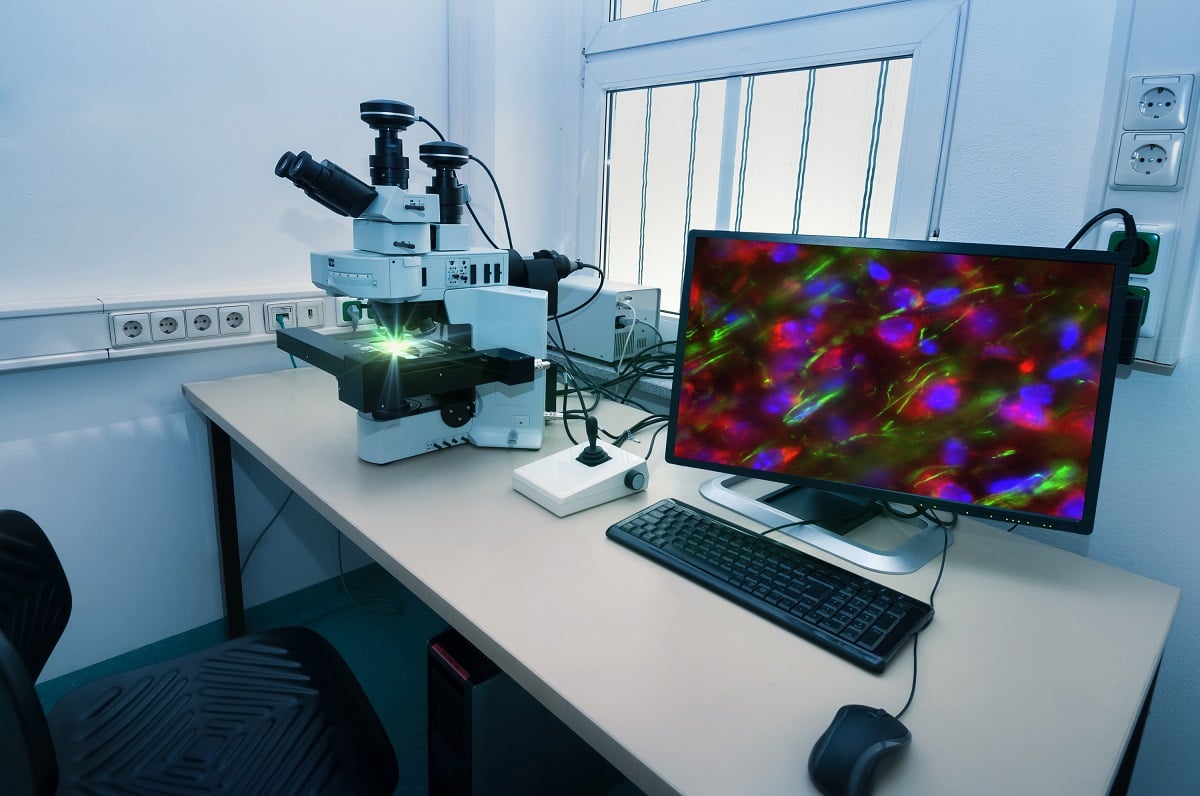 One of the miracles of modern science is the ability to use microscopy to view objects that cannot be seen with the naked eye. With microscopy, we are not limited to objects that are only able to be seen by the normal eye, which allows us to peer into worlds that even a few hundred years ago, scientists never imagined existed. The following is a description of thin section microscopy as well as ultra-thin section microscopy and how both affect and aid scientific research and discoveries.
Types of Microscopy
Understanding thin section or polished thin sections microscopy requires that an understanding of microscopy be attained. in its most simplest, microscopy utilizes micrscopes to view aspects of samples that we would have no idea existed without a more powerful method of viewing them. There are three well-known branches of microscopy: Optical, electron and scanning probe. With thin section preparation, a lab sample is provided using a polarizing petrographic microscope, electron microscope or electron microprobe. Each allows for visual penetration beyond what we would normally see with our unaided eyes or even with a traditional, scientific microscope.
Thin Section
To attain the proper sample, a thin section preparation service cuts a tiny portion of a rock, bone, wood or other substance and grinds it until it is optically flat. The sample is cut with a diamond saw to ensure the cleanest, quickest cut possible without degrading the sample through scorching or pulverizing. Once the sample is cut, personnel from thin section services then mounts the sample on a glass slide and the sample is then ground further using progressively finer abrasive grit. This process is completed when the sample is 30 micrometers thick. To verify the proper thickness, the sample is compared to an interference color chart that usually will use quartz as a gauge to determine thickness at that level because of quartz’s abundance as a mineral.
How Color is Used
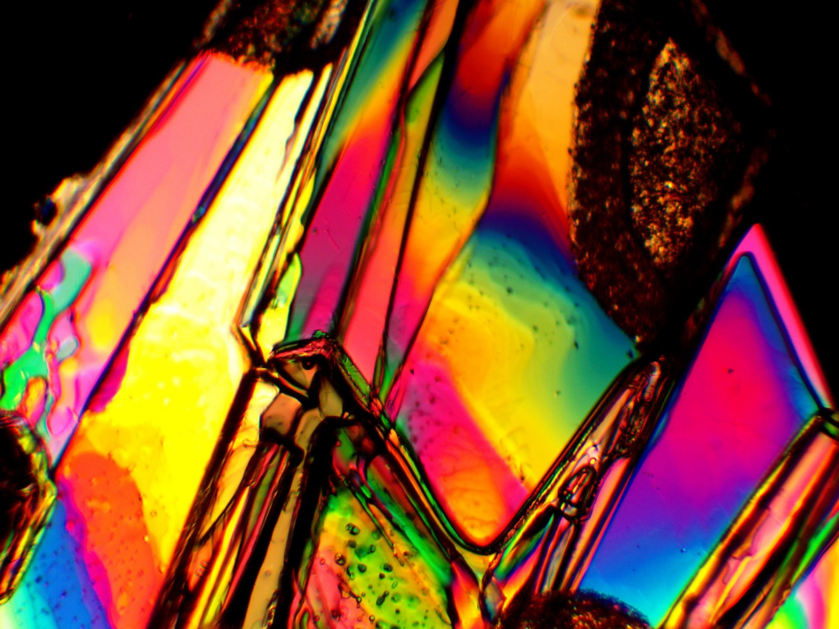
The thin section sample contains optical properties that are unique to each type of mineral or substance. When the sample is subjected to two polarizing filters set at right angles, those colors are altered as they are seen by the viewer. Because different minerals have different properties, minerals are able to be easily identified, based on the color they put forth. The color of a mineral is not as unique as a fingerprint because of the range of color, but it is unique enough for a positive identification of mineral presence in a sample.
 Why It Matters
Using microscopy and thin section sampling the optical properties of a sample are able to be investigated. Mineral properties in the sample are able to be isolated and identified. In this manner, the petrological process helps the scientist to discover the origin and evolution of the parent rock. This can aid in determining better classifications of rocks and minerals as well as helping to authenticate the history of a rock sample. Better classifications help us learn about the process rocks and minerals have gon through to get to the point they are at when they are ground for samples. Understanding the origin and history of a rock helps us better understand how it plays a role in the history of the earth itself.
Ultra-Thin Samples
In some samples, ultra-thin sections are prepared. Typically, samples that undergo this contain minerals of high birefringence. One example of that is the mineral calcite. In the process of attaining an ultra-thin sample, the 30-micrometer sample is prepared like a regular sample, but is attached to the slide using a soluble cement. This allows the sample to be worked on from two sides with a fine diamond paste to reduce the sample’s thickness. When the thickness reaches 2 - 12 micrometers, it is completed.
Practical Results
The ultra-thin method has been utilized to help prepare samples for transmission electron microscopy as well as to study the microstructures of minerals and rocks. Understanding microstructures helps us better understand how a rock was formed and how it has gotten to its current form, what stresses have helped form it and how its composition can affect an overall assessment of a sample. It also can help us with archeological studies as it lets us better understand the composition of manmade materials, such as concrete. This can shed light on the sophistication of a civilization as well as their building methods.
 Thin section sampling is the process by which a larger sample is reduced to the point it can be examined in the minutest detail, using color as a reference. Use this guide as a reference to understand microscopy, thin section and ultra-thin section samples as well as how they aid us in discovering our world.
