

Tools and Equipment Used in Palynology Labs
Palynology is the study of pollen and spores (collectively termed palynomorphs): tiny particles that provide crucial information in many fields. Analysis of modern and fossilized palynomorphs lends helpful data to locate oil and natural gas, determine the ages of rock layers, study allergies, identify plants used in honey, discover the diets of ancient civilizations. Palynology lab services carefully process and study palynomorphs using advanced tools, equipment & chemicals.
From Sample to Slide
Palynology labs start with a sample material, usually a specified amount of soil. In order to study the pollen and spores inside, palynologists must first remove the extra organic and inorganic material.
First, the sample is crushed to a specified degree, depending on the material. Acids or bases dissolve unwanted material, leaving only pollen. This process is called acetolysis. Finally, staining solutions like acetocarmine are often added to create a clearer image under a microscope. Once the palynomorphs are ready, a very thin amount is placed on a glass slide. These slides can be used under a microscope and stored for a long time.
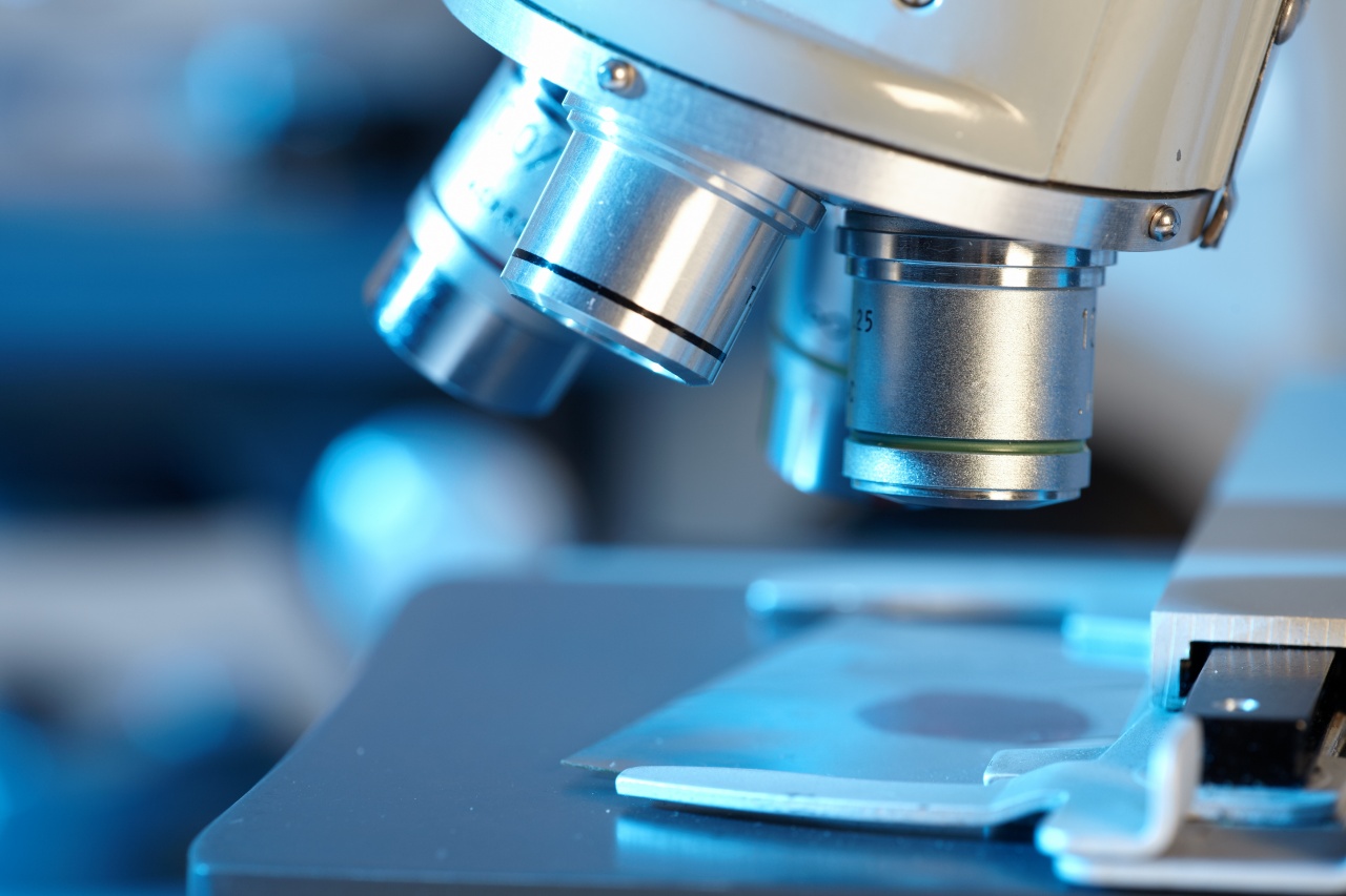
Microscopes
Most palynology labs use light microscopes and/or electron microscopes to gather details about pollen and spores. Light microscopes use light and lenses. They’re cheaper, smaller, and easier to use. However, light microscopes may not provide a resolution powerful enough to observe very small palynomorphs.
Electron microscopes use beams of electrons and electromagnets. They provide a much clearer and more magnified picture. Images are projected on a screen, so multiple palynologists can see the image at once. However, electron microscopes are more expensive and cumbersome than light microscopes.
| |


![]()
